ISSR CLASSES
Checkpoint Science
iGCSE Chemistry
iGCSE Physics
iGCSE Coordinated Science
A-Level Chemistry
PRACTICALS
Practicals Home
Practicals A-Z
Study Plan for Practical Work
The mass spectrometer is a machine which allows us to measure the mass of atoms and molecules with great accuracy. The following is a simplified diagram of one:
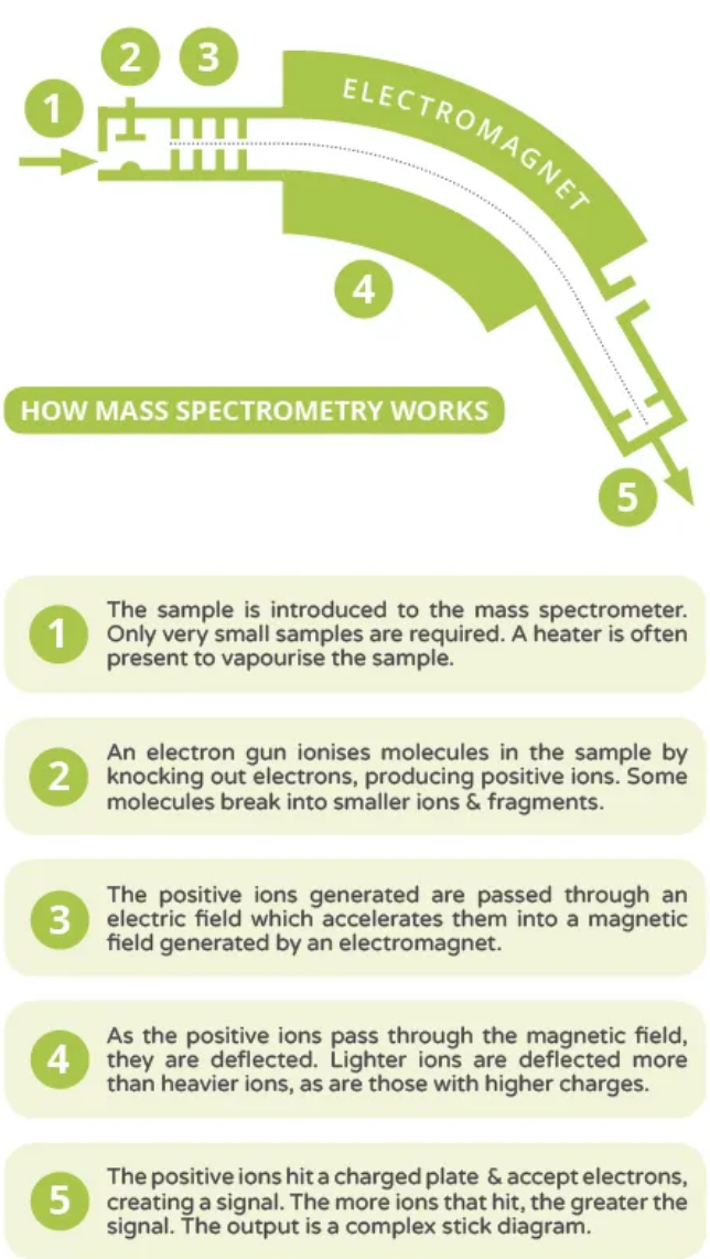
The air is removed from the apparatus by the vacuum pump, otherwise it would get in the way of the moving ions that are to be detected. Under the very low pressure of the apparatus the sample vaporises (substances boil at lower temperatures under reduced pressure) and the vapour is bombarded by high energy electrons from the electron gun. This causes electrons to be knocked out and positive ions are formed:
|
M(g) ⇒ M+(g) + e- |
Also, if the sample is made up of molecules, some of these will fragment - that is break up into smaller pieces. The various ions are accelerated towards the plate with the large negative potential and a fine beam of them streams through the narrow slit. These are then deflected (bent) by the strong magnetic field and some of them arrive at the detector and are recorded on a chart. Lighter particles are deflected more and heavier particles are deflected less, and so do not arrive at the detector. By adjusting the power to, and therefore the strength of the electromagnet, ions of different mass can be brought to the detector in turn and recorded. The chart shows the mass to charge ratio of the ion (m/e), although this can usually be considered to be the mass, as most of the ions have a charge of one. It also shows on the vertical axis the abundance (relative quantity) of all the ions present.
The analysis of the mass spectrum of a compound can be complicated. However, for "A" Level purposes a fairly simple method gives some good clues as to the identity of the compound. First look for the highest m/e value on the mass spectrum (it may not be the most abundant peak). This has been produced by the parent ion (molecular ion), that is the complete molecule with a 1+ charge. In real mass spectra this peak is not always detectable, but you should always find it in "A" Level problems. Real spectra are also complicated by the presence of isotopic peaks of higher mass, but these will have been removed from "A" Level exercises. The mass of the parent ion peak gives the relative molecular mass of the molecule.
|
The smaller mass peaks are the results of fragmentation, that is the parent ion falling apart under the extreme conditions present in the mass spectrometer. You will often be asked to suggest the formula for the ions which cause these fragment peaks. Just make sure they add up and the atoms are present in the original molecule! Under the very high energy conditions within the mass spectrometer the usual rules of bonding do not apply, so don't worry if you cannot see how your suggested fragment is held together. The presence of certain fragments gives us clues as to the structure of the original molecule. An m/e peak of 15 is caused by a methyl group, and one of 77 is caused by a phenyl group (C6H5) from an arene. |
|
More clues can be obtained by looking at the difference in mass between peaks. A peak appearing at 17 below the molecular ion mass suggests the loss of an OH group from the original molecule.
Mass spectra can be complicated by the presence of isotopes. These isotopic peaks and other complex details are often edited off problems set for "A" Level.
Example

The parent ion peak at 46 gives the molecule's molecular mass. The ion is C2H6O+
Loss of the OH hydrogen atom (-1) produces C2H5O+ at 45
Loss of a methyl group (-15) gives CH2OH+ at 31 (the most abundant peak, so most stable ion)
C2H5+ is seen at 29 (loss of OH, -17) and C2H3+ at 27 (loss of a further H2)
A small CH3+ peak is seen at 15
You can find some example problems in the spectroscopic analysis section.